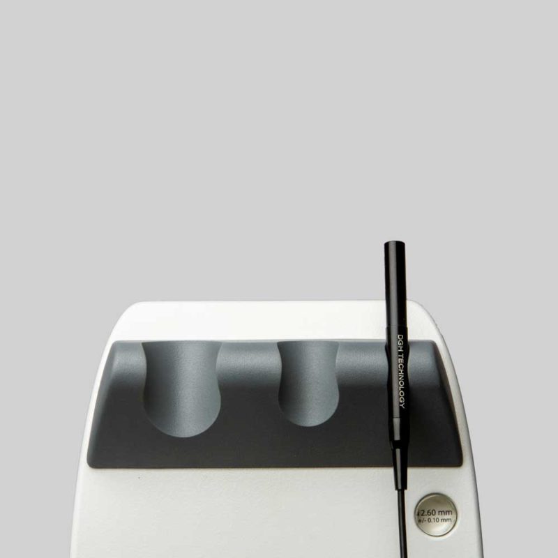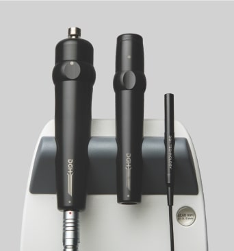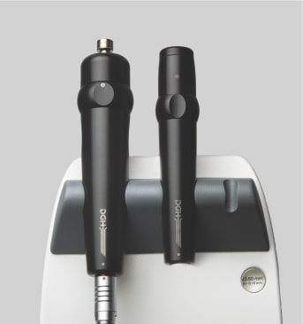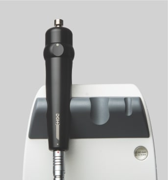
A-Scan
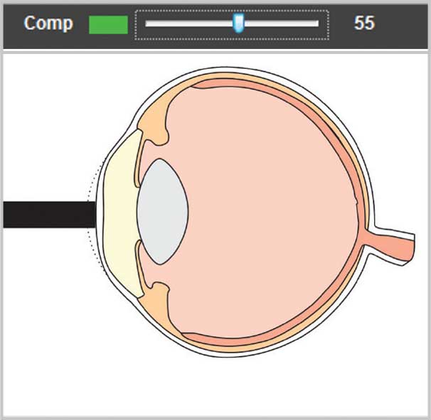
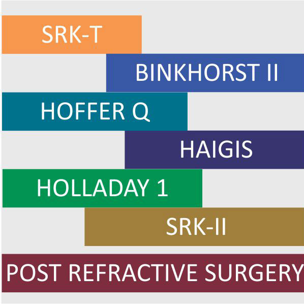
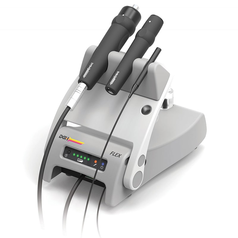
The Flex A-Scan uses DGH’s proven alignment algorithm to obtain repeatable and accurate axial length measurements. The IOL calculator is easy to use and includes most modern and post refractive formulas.
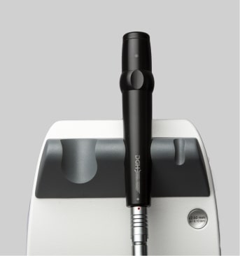
B-Scan
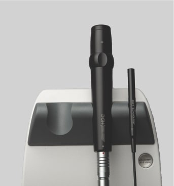
The redesigned B-Scan Probe, new with the Flex, provides clear imaging of the posterior segment of the eye, even when optical clarity is compromised.
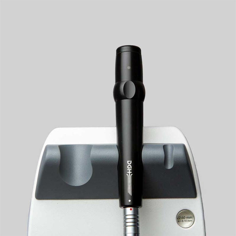
UBM
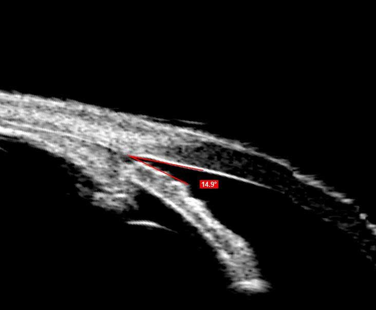
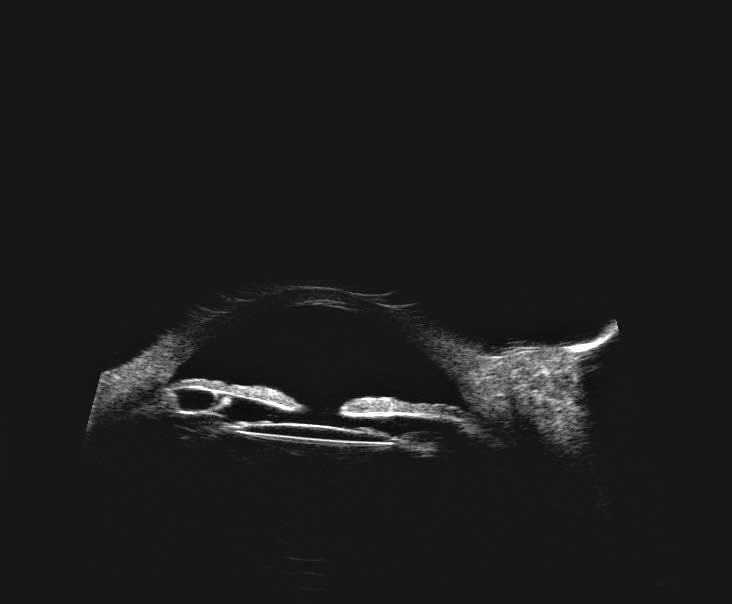
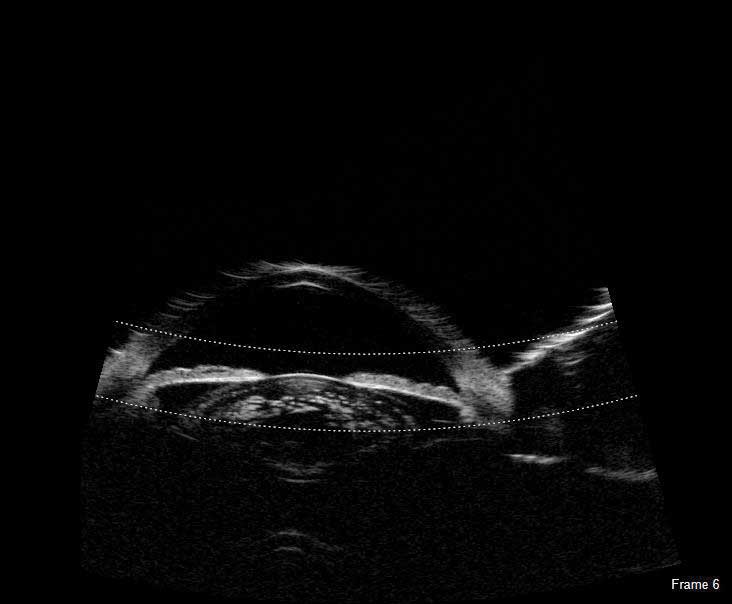
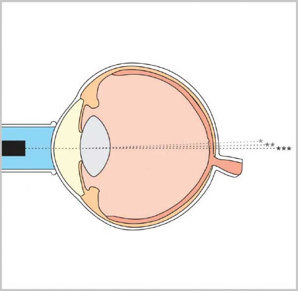
The Flex UBM probe is indispensable when obtaining high resolution images of the anterior segment of the eye, including images of structures concealed by the iris or corneal opacities.
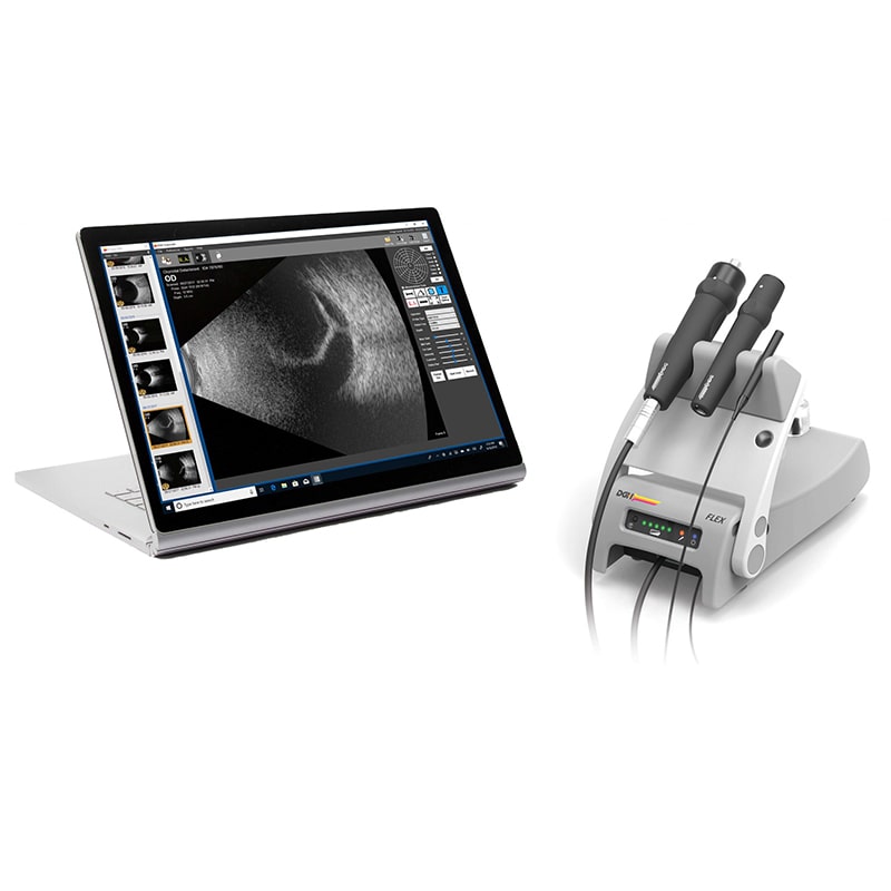
TOTAL IMAGING SOLUTION
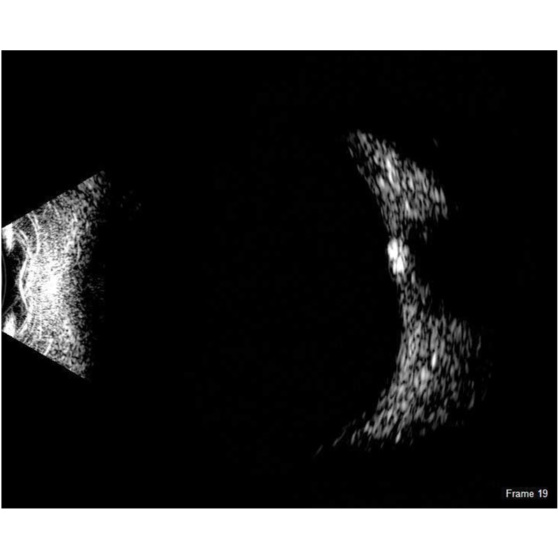
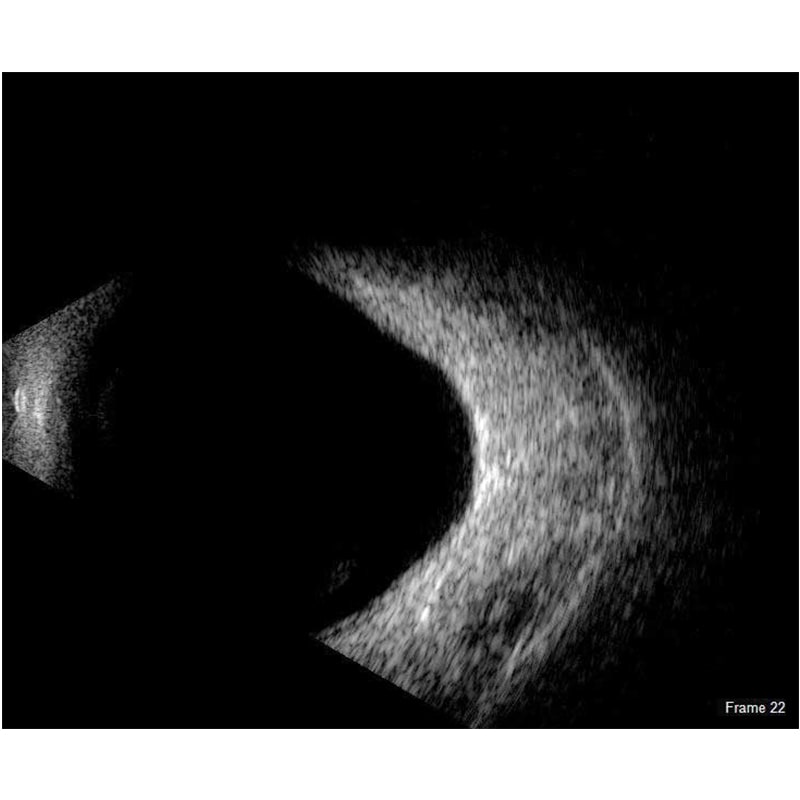
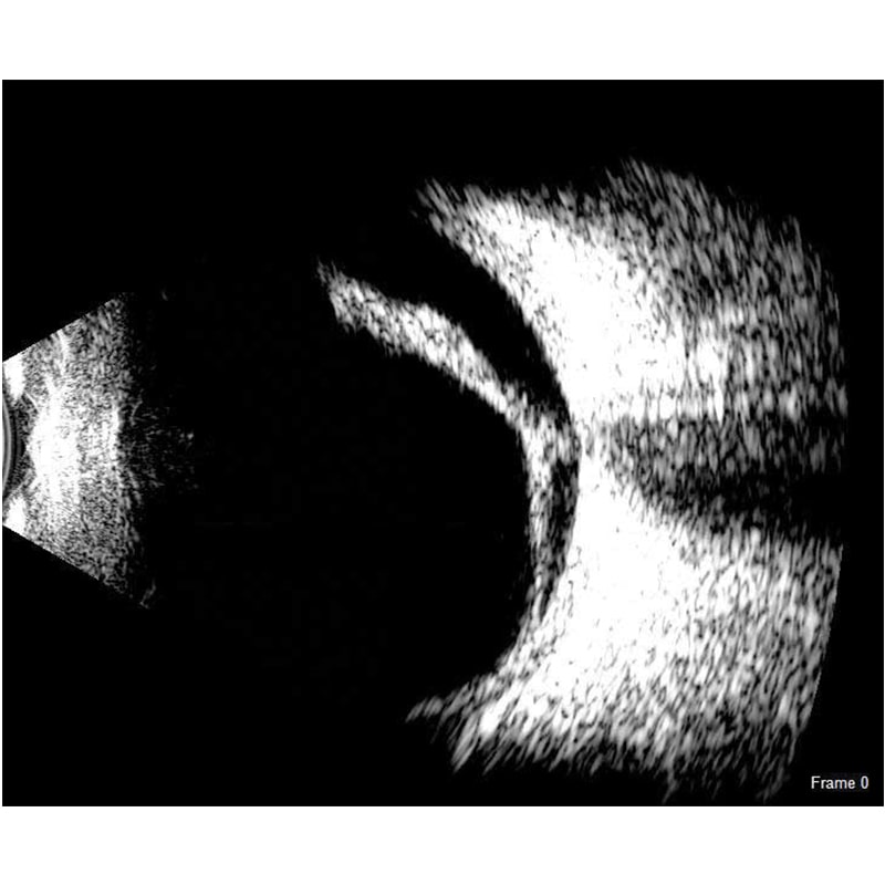
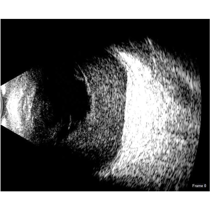
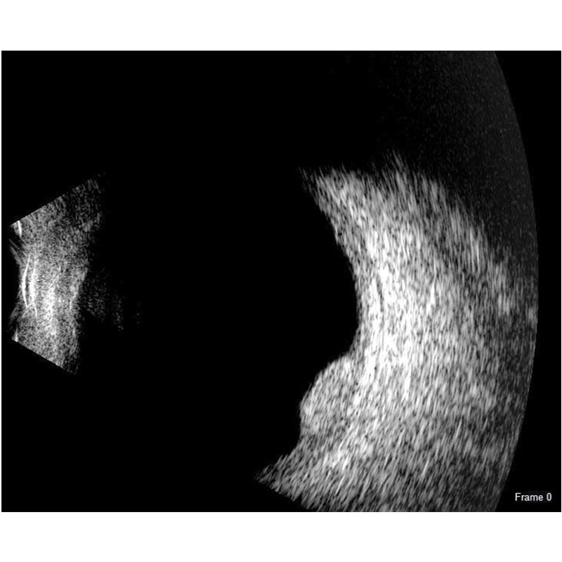
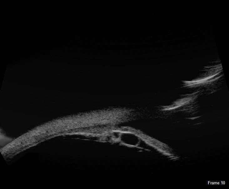
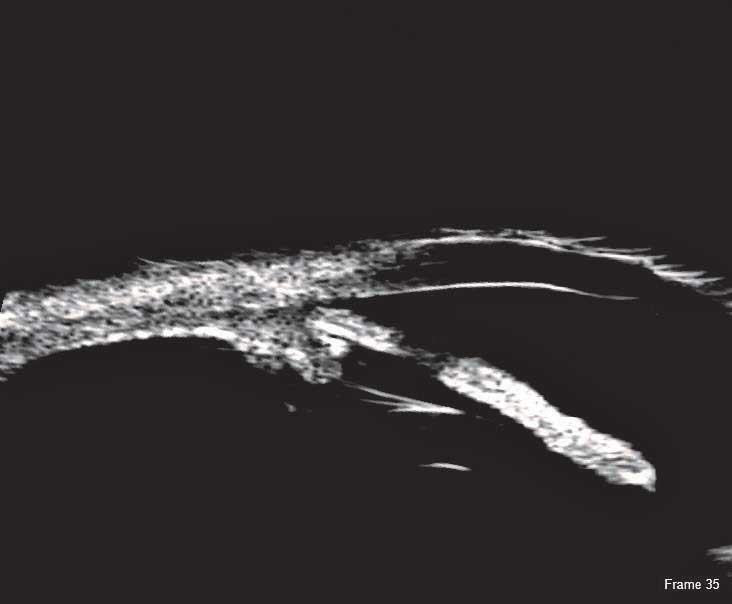
The Scanmate Flex B-Scan probe enables clinicians to capture clear and precise images and videos of the posterior segment of the eye. Ultrasonic B-scans are effective, even when opacities (such as dense cataract, blood, or anatomical structures) are present which obscure optical technologies.
The B-Scan probe is available in both 12.5 MHz and 20 MHz frequencies. Among the on-screen tools are calipers to measure structures, an area measurement tool and an annotation tool that gives you a way to indicate pathologies on the image.tool and an annotation tool that gives you a way to indicate pathologies on the image.
B-Scan Diagnostic Applications
The Flex B-Scan delivers clear images of the posterior portion, even when optical clarity is compromised. B-Scan imaging can aid the evaluation of:
- Retinal Detachments
- Vitreous Detachments
- Vitreous Humor Pathologies
- Staphylomas
- Posterior Segment Pathologies
- Choroidal Pathologies
- Optic Nerve Pathologies
- Scleral Thickening