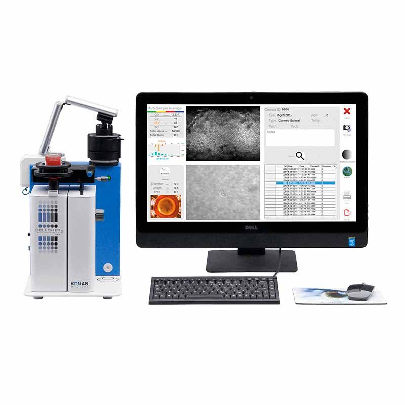
CellChek D and EB-10 Specifications
| CellChek D+ | EB-10 |
|---|
| Viewing Field | 1000 x 750 µm | 480 X 600 µm |
| Analysis Area (each multi-field area) | 400 X 300 µm X 4 areas
(max 480,000 µm2) | 200 X 280 µm |
| Chamber Compatibility | Numedis Life 4C®, Krolman®, B+L®, Stephens® | Life 4C®, Alcon®, Bausch+Lomb®, Krolman® |
| Vial Dimensions | up to 35 mm diameter | up to 35 mm diameter |
| Thermometer Range | 0° to 45° C | 0° to 45° C |
| Stage Translation Ranges | X, Y, and Z = 16 mm, Tilt = 15° | X and Y = 16 mm, Z= 20 mm, Tilt: 5° |
| Illumination | Halogen | LED: primary wavelength 525 nm |
| Cameras | Dual CMOS: Finder and cornea | Dual CMOS: Finder and Cornea |
| Display | Digital data feed to computer | On-board LCD: temperature and pachymetry |
| Electrical | 100-240 VAC, 50/60 Hz, 50 VA | 100-240 VAC, 50/60 Hz, 50 VA |
| Size | 280 (W) X 215 (H) X 265 (D) mm | 200 (W) X 255 (H) X 220 (D) mm |
| Data Interface | USB 2.0 | USB 2.0 |
| Weight | 7.5 kg (without computer) | 6.1 kg (without computer) |
| Operating Conditions | Ambient temp: 10° to 40° C
Relative humidity: 30% to 85%
Atmospheric pressure: 70 to 106 kPa | Ambient temp: 10° to 40° C
Relative humidity: 30% to 85%
Atmospheric pressure: 70 to 106 kPa |
| Regulatory | CE Mark, In Vitro USA | CE Mark, In Vitro USA |
What is KonanCare support and clinical training?
KonanCare enables customers to consult, securely, by appointment, with Konan’s on-staff experts for interpretation of specular microscopic images, and to receive assistance on more advanced topics and methods associated with Konan’s products (CellChek XL, EvokeDx, CellChek D+ and EB-10, Chart2020, and the RAPDx pupillograph). With current product models, a secure, permission-based login session can be initiated by the customer allowing both the physician and the KonanCare team member to simultaneously view and discuss actual images using secure internet web services. KonanCare assistance is provided without charge for customers during the initial warranty period or during any period of Service Agreement coverage. A unique service, only from Konan Medical USA.
How accurate are the analysis methods?
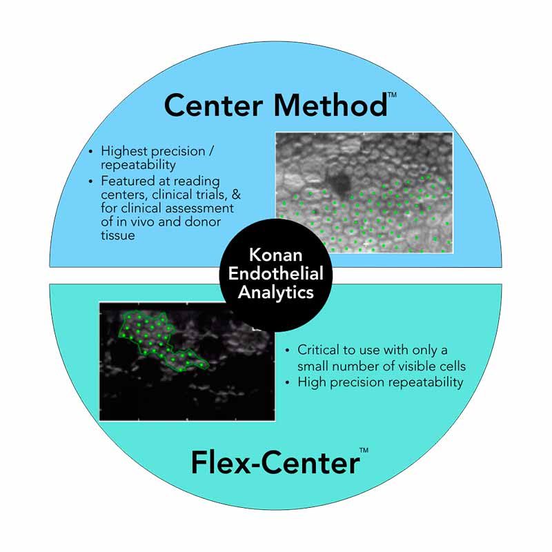
Flex Center MethodKonan Center/Flex Center methods are patented and are the only FDA 510(k) cleared methods for eye bank systems. Accuracy established by independent reading centers for FDA clinical studies is +/-2.5%
How is the system calibrated?
Each CellChek D+ comes with a 20um grid calibration disk. Enter 100um length using this disk and click a button. The computer will indicate a known magnification for 100um length. It also enables the operator to verify if calibration was properly completed.
How many cells should be counted to be statistically accurate?
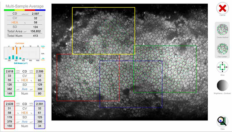
If the cells in the sample area are generally uniform in size and shape, 30-50 cells should be enough. However, if there is variation in cell size, at least 75-100 cells should be counted. Konan’s multi-sampling capability enables the operator to choose up to 4 different locations in the central cornea for adequate sampling. Analysis from each frame can be automatically averaged.
How accurate is the built-in pachymeter?
CellChek D+ features improved visualization of both endothelial and epithelial surfaces, but has not yet been independently assessed as to the accuracy of the pachymeter. The previous model EB-10 has accuracy of +/-10%, however, accuracy in the donor cornea is dependent upon how well the epithelium is preserved.
How do I set a focal distance between the objective lens and the cornea placed in different containers, such as vial or chamber?
The operator is able to view the proper height of the stage by using the focusing knob and placing a slit of light reflection from the endothelium in the middle of a green box.
What temperature range is required for corneal assessment (after removing from refrigeration)?
CellChek D+ has a built-in thermometer which measures medium temperature in real-time. For optimal results, the preservation medium should be 24 – 26 degrees C.
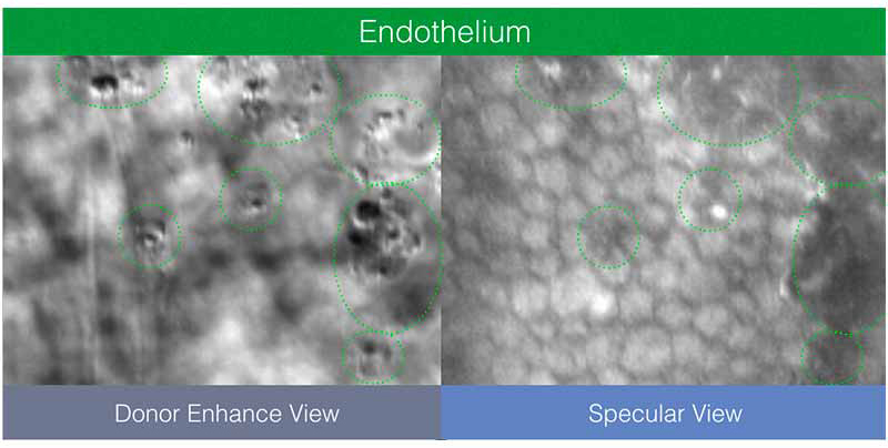
How is the imaging affected with cold corneas and how do we determine if the cornea has warmed up enough to analyze?
At 4º C, biochemical and physiological metabolic activities of endothelial cells slow down significantly. As temperature is increased to ~ room temperature, (>24º C), pumping mechanism is reactivated. If cornea has low functional reserve, it may take longer for activation to occur.