
CIRRUS™ with AngioPlex™ creates a new era in both OCT and angiography.
For the first time, AngioPlex allows for the visualization of both vascular and structural information from a single non-invasive scan. One that makes eye care’s leading clinical OCT platform an unprecedented tool for the acquisition of ultra-clear color-depth-resolved 3D microvascular imaging of the retina. AngioPlex technology revolutionizes clinical practice by making the visualization of microvasculature of the retina a routine part of everyday care.
New vascular information
Ultra-clear 3D microvascular visualizations powered by OMAGc
OMAGc detects motion of red blood cells within sequential OCT B-scans performed repeatedly at the same location
Depth of retinal vasculature color-coded for ease of visual assessment
Advancing Smart OCT
Enter the New Era of Retinal Care
Now, ultra-clear 3D microvascular visualizations with non-invasive technology can be a routine part of your retina practice
New Vascular Information
Depth–resolved visualization of the retinal vasculature. Powered by Optical Micro Angiography (OMAGc) Algorithms that utilize amplitude and phase OCT signal data to deliver the highest-quality ultra-clear 3D angiography images.
Improved Workflow
FastTrac™ provides live-tracking for motion-artifact-free images. Single-Scan Simplicity ensures ZEISS AngioPlex requires only a single additional OCT scan to generate an ultra-clear 3D OCT angiography image.
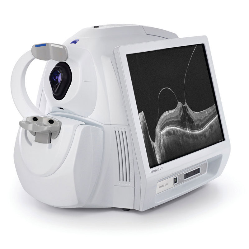
Most Powerful OCT Platform in the Market
AngioPlex OCT Angiography is available on the CIRRUS 5000 HD-OCT platform, allowing ophthalmic practices the flexibility to easily integrate vascular imaging with standard OCT diagnostic imaging.
Clinical Cases
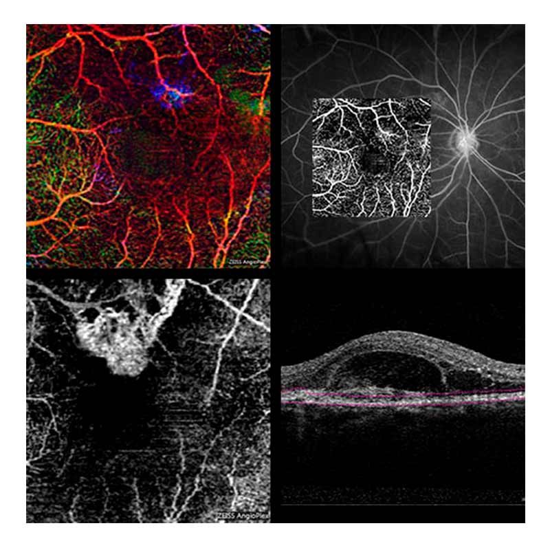
Diabetic Retinopathy (DR) (Clockwise from top left)
AngioPlex images can illustrate the presence of microaneurysms and areas of ischemia
- Full depth color encoded image
- Superficial Retinal Layer
- Deep Retinal Layer
- Superficial layer overlaid onto FA
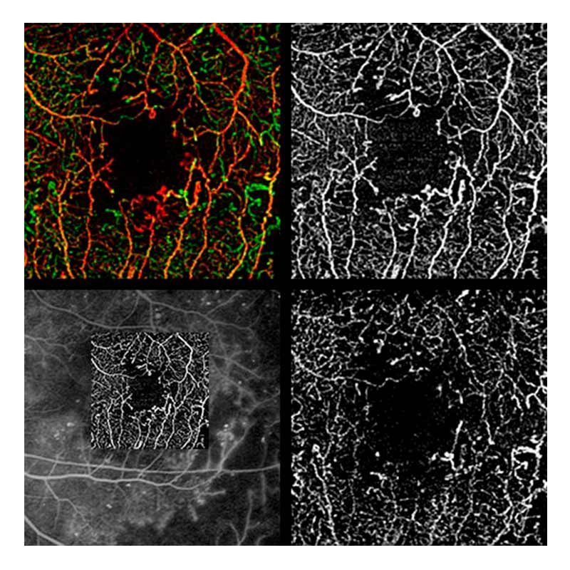
Age related Macular Degeneration (AMD) (Clockwise from top left)
AngioPlex images beautifully illustrate the presence of choroidal neovascularization (CNV)
- Full depth color encoded image
- Superficial Retinal Layer overlaid onto FA
- B-Scan
- Custom layer revealing CNV below the RPE
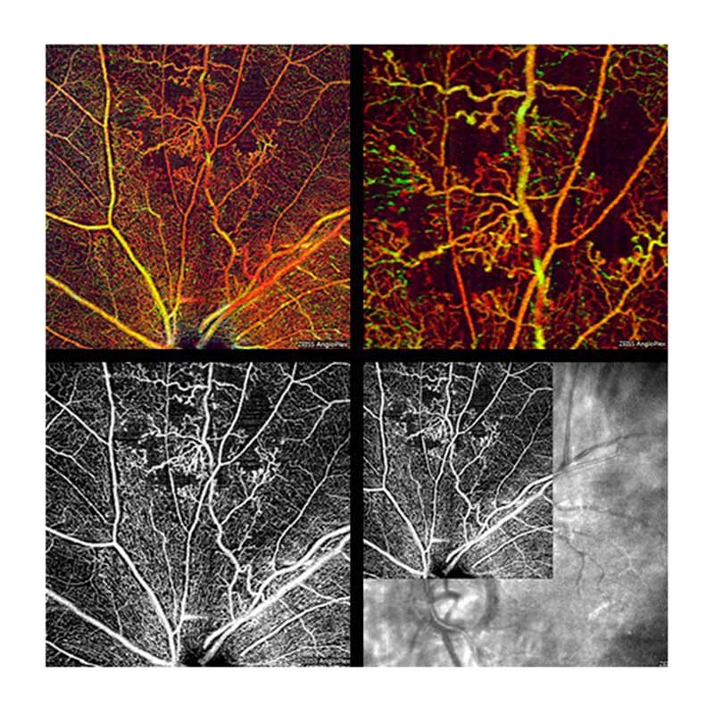
Branch Retinal Vein Occlusion (BRVO) (Clockwise from top left)
AngioPlex images clearly delineate the location of the occlusion and affected areas of ischemia superior to optic nerve head
Full depth color encoded image, 6x6mm image
Full depth color encoded image, 3x3mm image
Superficial retina layer overlaid onto LSO Fundus image
Superficial retina Layer