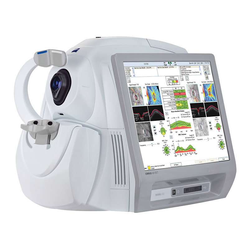
The Essential OCT CIRRUS HD-OCT 500
Offers comprehensive care practices essential OCT capabilities with a broad range of clinical applications in an easy-to-learn, easy-to-use instrument. It aids in the management of glaucoma and retinal disease, retina assessment for cataract surgery and anterior segment imaging for corneal disease.
Brilliant Visualizations
Examine Retinal Details: Selective Pixel Profiling™ optimizes each illumination point in the 20 X HD Raster Scan, ensuring detail-rich visualizations that spotlight critical pathological elements.
Gain Deeper Insight: New Enhanced Depth Imaging focuses the signal lower in the scan window for assessment of the deeper choroidal tissue.
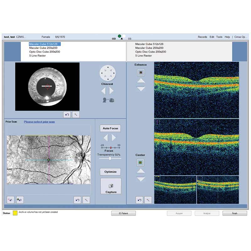
Visualize Change: New FastTrac™ on the CIRRUS HD-OCT 5000 precisely targets and captures the same tissue every time to ensure consistent comparison.
Certainty at the speed of CIRRUS
Insightful Analyses
Reproducible Analyses: Zeiss propietary algorithms measure and display layers for unsurpassed tissue targeting, segmentation, and reproducible measurements.
Comparative References: Diversified normative databases of ONH, RNFL, Ganlion Cell / IRL, and Macular Thickness facilitate at-a-glance identification of anatomy outside normal limits.
Change Measurements: All Cirrus data cubes are automatically registered with historical data, allowing for point-to-point measurements of change.
Software Version 6.5 Features for Cirrus 500
Advanced diagnostic tools that improve your ability to identify pathology and track change over time.
NEW Macular Thickness OU Analysis
Ganglion Cell Analysis
Guided Progression Analysis (GPA™)
Precision FoveaFinder™
Macular Thickness and Change Analysis
Great Images & Dense Data Cubes
Analyze one pathology from multiple views!
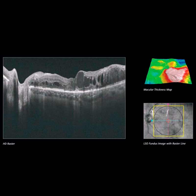
Cirrus Enhanced HD Raster Scans are second to none. Capture highly dense data cubes for all the detail you need for analyses and B-scans that help identify even the most subtle pathology.
Enhanced High Definition Scans
With With world-class ZEISS optics and knowledge from over a decade of experience in OCT, Cirrus captures spectacular images second to none. Rather than image averaging, Cirrus uses Selective Pixel Profiling™ to optimize data at each pixel. Judge the results for yourself.
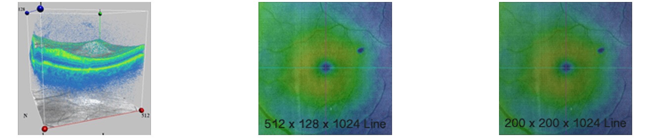
| Scan Pattern | Data Points per A-scan | Total Data Points | Spacing between lines | Capture time |
| 512 X 128 | 1024 | >67 million | 47 µm | 2.4 s |
| 200 X 200 | 1024 | >40 million | 30 µm | 1.5 s |
The Power of the Cube Cirrus captures a highly dense cube over a 6 mm x 6 mm area. The cube data is used for both visualization and analysis; no additional scan pattern is needed for analysis. More than 67 million data points!
Why Should You Care?
Spot small areas of pathology. Tightly spaced B-scans, either 30 or 47 microns apart in the cube, ensure that small areas of pathology are imaged. For reference, a human hair is about 40-120 microns in diameter.
Visualize the fovea. Scans that are spaced further apart than in the Cirrus cube may miss the central fovea.
Fuel for analysis. Millions of data points from the cube are fed into the ZEISS proprietary algorithms for accurate segmentation, reproducible measurements and registration for change analysis.
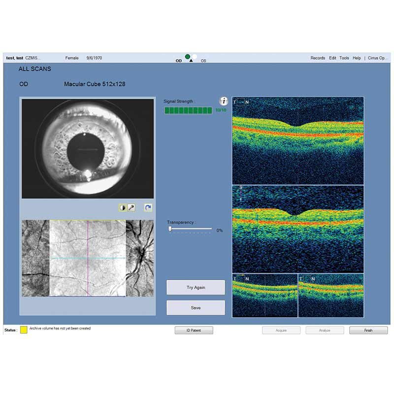
Take the pressure off the operator. As long as the scan is roughly placed on the fovea or optic nerve, the software automatically centers the measurements after the capture.
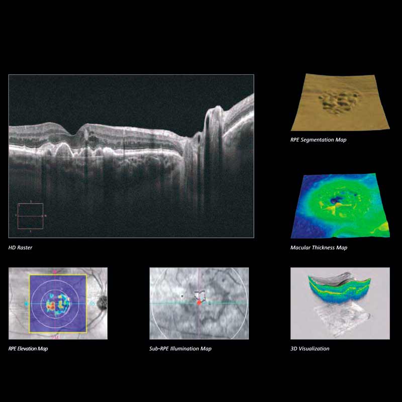
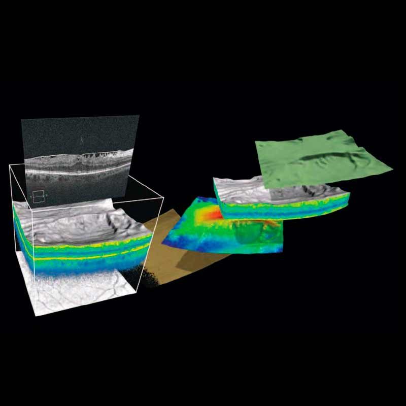
See the tissue from different perspectives. View the cube data from all angles, with 3D rendering, OCT fundus images and customized en face slabs.
3D and Advanced Visualization™
Draw insights from perspectives beyond the cross-sectional B-scan. Cirrus data cubes can be processed and mined with sophisticated 3D and en face visualization tools. Isolate layers of the retina to view en face. Fly through from different angles. The answers are in the cube and Cirrus provides the tools to find them.
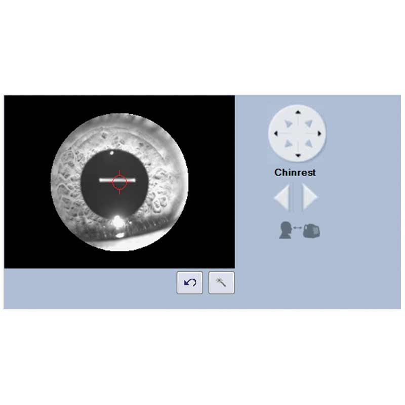
Anterior Segment Images
Cirrus can image the angle or cornea with raster scans or a data cube. No add-on lens is required.
Age-Related Macular Degeneration