
ZEISS AngioPlex OCT Angiography
The introduction of ZEISS AngioPlex ushered a new era in retinal care.
Now, ultra-clear 3D microvascular visualizations with non-invasive technology can be a routine part of your retina practice.
AngioPlex OCT-A allows you to visualize without the use of a contrast agent.
Advancing Smart OCT for Anterior Segment, Glaucoma and Retina
Anterior Segment Premier Module
PanoMap Wide-Field Display
Smart HD Scans
En face report
Advancing Smart OCT
Visualization at the speed of CIRRUS
Analyzing a single pathology from multiple views provides comprehensive insight and analysis of the clinical situation. How this helps you:
Spot small areas of pathology. Tightly spaced B-scans, (either 30 or 47 μm apart), in the cube ensure that small areas of pathology are imaged. For reference, a human hair is about 40-120 μm in diameter.
Visualize the fovea. Scans that are spaced further apart than in the CIRRUS cube may miss the central fovea.
Fuel for analysis. Millions of data points from the cube are fed into the Zeiss proprietary algorithms for accurate segmentation, reproducible measurements and registration for change analysis.
Take the pressure off the operator. As long as the scan is placed in the vicinity of the fovea or optic nerve, the software automatically centers the measurements after the capture.
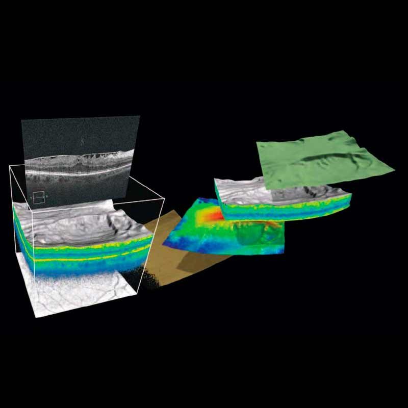
See the tissue from different perspectives. View the cube data from all angles, with 3D rendering, OCT fundus images and Advanced Visualization™.
Future ready. Previously captured CIRRUS cubes can be analyzed using new analyses.
Tracking at the speed of CIRRUS
FastTrac™ reduces eye motion artifacts without sacrificing patient throughput with a proprietary scan acquisition strategy, high speed 20 Hz LSO camera, and single-pass alignment scanning. With FastTrac, scan at the highest resolution at the same location at each visit.
Assessment at the speed of CIRRUS
Measurement centering with FoveaFinder and AutoCenter
On the macula, the unique FoveaFinder technology ensures the ETDRS and ganglion cell plus inner plexiform layer measurement frameworks are centered on the fovea.
AutoCenter™ function automatically centers the 3.4 mm diameter peripapillary RNFL calculation circle around the disc for precise placement and repeatable registration. The placement of the circle is not operator dependent. Accuracy, registration and reproducibility are assured.
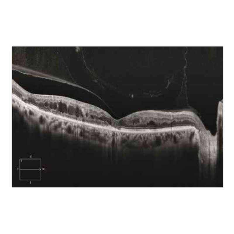
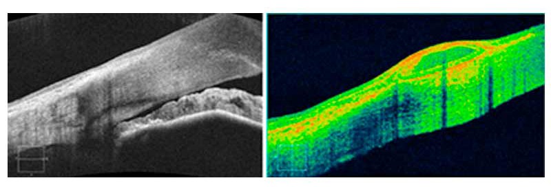
New Smart HD 1 Line scan
Captures and averages 100 b-scan
images with automatic centering
at the fovea or region of interest.
The result is a brilliant image that
simultaneously highlights detail in
the vitreous, retina, and choroid.
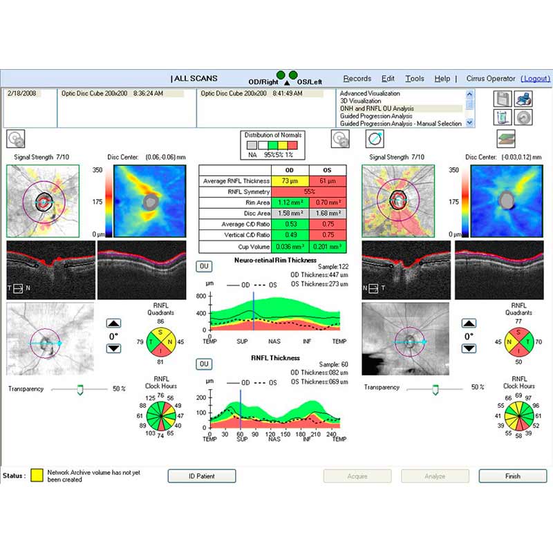
Assessment at the speed of CIRRUS
Measurement centering with FoveaFinder and AutoCenter
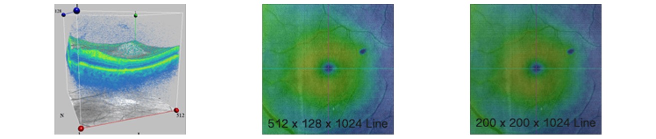
Great Images & Dense Data Cubes
Analyze one pathology from multiple views!
Cirrus Enhanced HD Raster Scans are second to none. Capture highly dense data cubes for all the detail you need for analyses and B-scans that help identify even the most subtle pathology.
Enhanced High Definition Scans
With world-class ZEISS optics and knowledge from over a decade of experience in OCT, Cirrus captures spectacular images second to none. Rather than image averaging, Cirrus uses Selective Pixel Profiling™ to optimize data at each pixel. Judge the results for yourself.
| Scan Pattern | Data Points per A-scan | Total Data Points | Spacing between lines | Capture time |
| 512 X 128 | 1024 | >67 million | 47 µm | 2.4 s |
| 200 X 200 | 1024 | >40 million | 30 µm | 1.5 s |
The Power of the Cube Cirrus captures a highly dense cube over a 6 mm x 6 mm area. The cube data is used for both visualization and analysis; no additional scan pattern is needed for analysis. More than 67 million data points!
Why Should You Care?
Spot small areas of pathology. Tightly spaced B-scans, either 30 or 47 microns apart in the cube, ensure that small areas of pathology are imaged. For reference, a human hair is about 40-120 microns in diameter.
Visualize the fovea. Scans that are spaced further apart than in the Cirrus cube may miss the central fovea.
Fuel for analysis. Millions of data points from the cube are fed into the ZEISS proprietary algorithms for accurate segmentation, reproducible measurements and registration for change analysis.
Take the pressure off the operator. As long as the scan is roughly placed on the fovea or optic nerve, the software automatically centers the measurements after the capture.
See the tissue from different perspectives. View the cube data from all angles, with 3D rendering, OCT fundus images and customized en face slabs.
3D and Advanced Visualization™
Draw insights from perspectives beyond the cross-sectional B-scan. Cirrus data cubes can be processed and mined with sophisticated 3D and en face visualization tools. Isolate layers of the retina to view en face. Fly through from different angles. The answers are in the cube and Cirrus provides the tools to find them.
Anterior Segment Images
Cirrus can image the angle or cornea with raster scans or a data cube. No add-on lens is required.
Cirrus HD-OCT Models 5000 and 500 Specifications
Third Party Software and Hardware
The following hardware and software is compatible with the CIRRUS ™ HD-OCT:
Virus Scanning
The CIRRUS HD-OCT Software has been tested with Microsoft® Security Essentials, but anti-virus software does not come pre-installed. When installing your anti-virus software per the manufacturer’s recommendations, configure it so it does not scan automatically, as this may interrupt the operation of the CIRRUS instrument or your review station. Virus scans and virus definition updates must be launched manually or scheduled to run when patient data acquisitions are not actively being performed on the CIRRUS equipment.
Operating Systems
Software Compatibility
- CIRRUS Version 7.6/8.1 Software: Windows 8.1 (32- and 64-bit), Windows 7 SP1 (32- and 64-bit), Windows Server 2008 R2, and Windows Server 2012 R2.
- CIRRUS Version 9.0 (and higher): Windows 8.1 (64-bit), Windows 7 SP1 (64-bit), Windows Server 2008 R2, and Windows Server 2012 R2.
Restrictions: Refer to Software Installation Instructions for minimum system requirements.
Compatible Printers
- Brother HL-2700CN*
- EPSON WorkForce 60
- HP 3520
- HP 6000*
- HP 6980*
- HP 6988*
- Samsung CLP-415NW**
- Samsung C1810W**
- Postscript Compatible printers**
*Only approved for 4000/400.
**Must use generic Postscript printer driver pre-installed on instrument
Restrictions:
- Device must be outside the patient environment (at least 1.5 meters away), unless used with an isolation transformer.
- Do not use with an extension cord or power strip.
- Use unshielded network cables (UTP) only.
- Do not share the same wall outlet with the CIRRUS HD-OCT.
- Contact us for compatible printer driver version(s).
Network Switches
- Interface speed: 10/100/1000 Mb/s
- Interface connectors: RJ-45
- Cable detection: MDI/MDI-X (automatic straight-through or crossover cable detection)
- Supported standards: IEEE 802.3, 802.3u, 802.3ab, and 802.3x
Restrictions:
- Device must be CD-Marked for use in EU
- Device must have Nationally Recognized Testing Laboratory (NRTL) and FCC approval for use in the US.
- Device must be powered by USB port only.
- Device must be outside the patient environment (at least 1.5 meters away), unless used with an isolation transformer.
- Do not use with an extension cord or power strip.
- Use unshielded network cables (UTP) only.
- Do not share the same wall outlet with the CIRRUS HD-OCT.
Upgrades
Product and Software News
Current software releases for up-to-date functionality
Make sure your ZEISS device is up to date with the latest software release. Below are the current software versions recommended for your Zeiss instruments. Maintenance upgrades are free of charge*.
- Cirrus HD-OCT 5000…………….9.5**
- Cirrus HD-OCT 500……………..9.5
- Cirrus HD-OCT 4000 Quad Core……8.1
- Cirrus HD-OCT 4000 Dual Core……8.0
- Cirrus HD-OCT 400……………..7.5
A shipping and handling fee may apply to maintenance upgrades. ** Requires 64 bit computer for CIRRUS Review Software. Otherwise CIRRUS HD-OCT 8.1 software is recommended.
Cirrus Software Version 9.5 Features
- Anterior Segment Premier Module with Lens Kit
- Smart HD-Scans
- FastTrac™ retinal tracking system
- Macular Thickness OU Analysis
- Advanced RPE analysis
- Ganglion Cell Analysis
- Guided Progression Analysis (GPA™)
- Precision FoveaFinder™
- Macular Thickness and Change Analysis
- Macular Thickness Normative Data