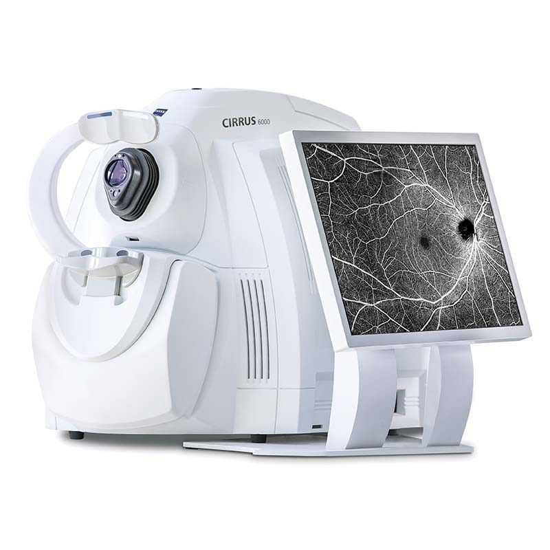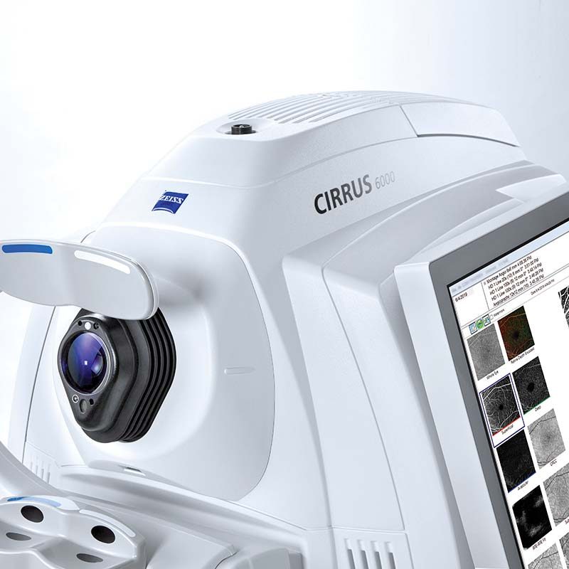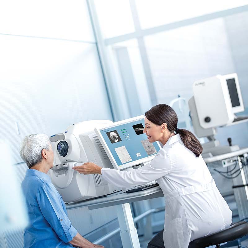
Performance OCT
Faster imaging with greater detail, at 100,000 scans per second, for improved patient care.
Proven analytics
Comprehensive, clinicallyvalidated tools to diagnose and manage a range of conditions.
Patient-first design
Seamless transfer of raw patient data from previous generations of CIRRUS for continuity of patient care.
The power of 100,000 scans per second
Faster imaging
Reduce chair time and speed up your practice.
• 270% faster OCT scans and 43% faster OCTA scans.*
• OCT cube scans in as little as 0.4 seconds.
• High-speed imaging in combination with FastTrac™ eye tracking technology reduces the chance of motion artifacts such as those caused by blinks and saccades.
Greater detail
View more in seconds and dig deeper with high-definition imaging.
• 12×12 mm single-shot OCTA cube scan in addition to 8×8, 6×6 and 3×3 mm scans.
• High-Definition AngioPlex scans (8×8 and 6×6 mm) for even greater microvascular detail without limiting the field of view.
• 2.9 mm scan depth.
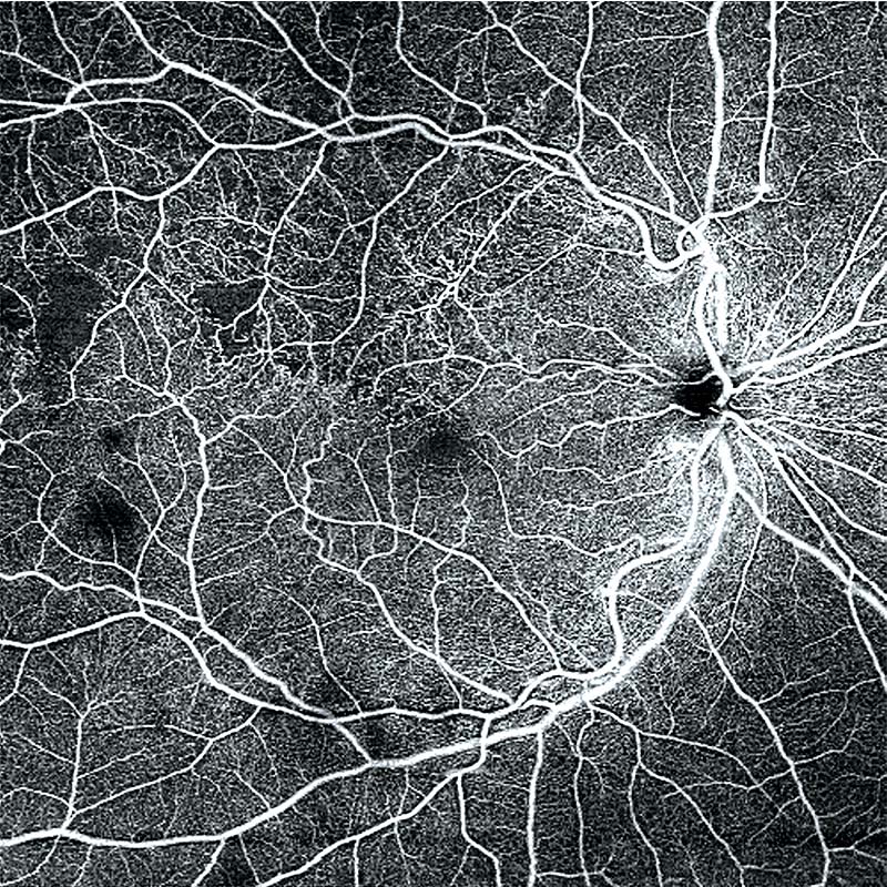
12×12 mm single-shot OCTA of branch retinal vein occlusion (BRVO).
Image courtesy of Jesse Jung, MD, East Bay Retina, United States
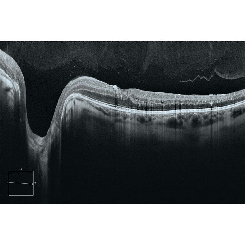
12 mm HD 1 Line Raster 100x averaged.
Image courtesy of Theodore Leng, MD, Byers Eye Institute, United States
Proven analytics
CIRRUS-powered treatment decisions
As the pioneering OCT technology, the CIRRUS platform offers clinicians extensive, clinically-validated applications for retina, glaucoma and anterior segment. The result: precise analysis, faster throughput and smarter decision-making across a wide spectrum of clinical conditions and patient types.
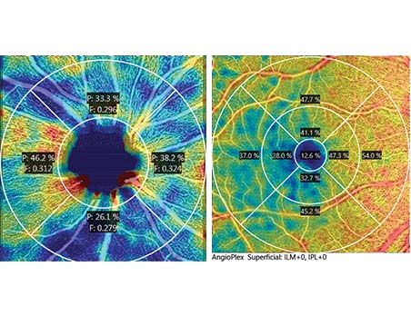
AngioPlex Metrix OCTA Quantification
AngioPlex® Metrix™ for Macula and ONH
AngioPlex Metrix allows clinicians to objectively assess and track progressive eye diseases such as diabetic retinopathy and glaucoma with quantification tools such as Vessel Density, Perfusion Density, and Foveal Avascular Zone (FAZ) for the macula, and Capillary Flux Index for the optic nerve head.
Patient-first design
Unique platform designed for the future
With ZEISS CIRRUS 6000, your patient data is never left behind. The CIRRUS platform ensures seamless transfer of raw, dynamic patient data from previous generations of the device – enabling clinicians to maintain continuity of patient care, even as OCT technology evolves over time.

Methodology | Spectral domain OCT |
Optical source | Superluminescent diode (SLD), 840 nm |
Scan speed | 100,000 scans per second |
A-scan depth | 2.0 – 2.9mm (in tissue) |
Axial resolution | 5 μm (in tissue) |
Transverse resolution | 15 μm (in tissue) |
Fixation | Internal, external |
Internal Fixation (focus adjustment) | -20D to +20D (diopters) |
Imaging Modes | Posterior segment, Anterior Segment, OCT Angiography, Fundus Imaging |
B-scan | 12 mm Raster; 3-12 mm cube |
OCTA | 3x3mm, 6x6mm, 8x8mm, 12x12mm (Retina); 4.5×4.5mm (Optic Nerve Head), 14×14 mm, 14×10 mm |
Instrument Weight | 35 kg (77 lbs) (without monitor) |
Instrument Dimensions (WxDxH) | 62.2L x 42.5W x 29.2H cm (24.4L x 16.7W x 11.4H in) |
Monitor | 22” Widescreen HD |
Input devices | wireless keyboard, wireless mouse |
Operating system (Instrument) | Windows® 10 |
Processor | i7 Intel® processor (7th gen) |
