
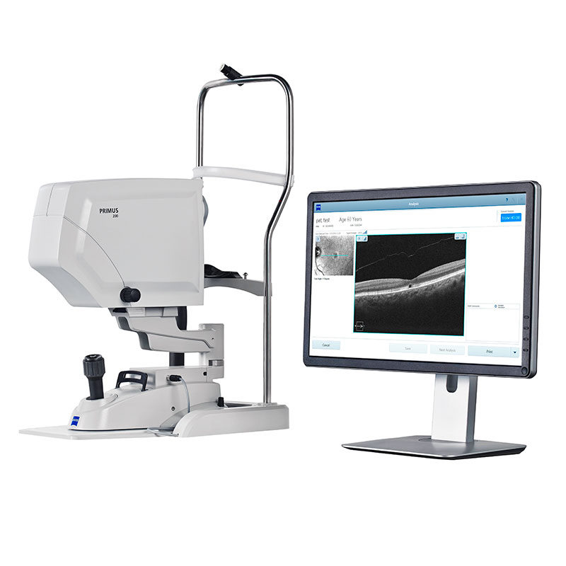
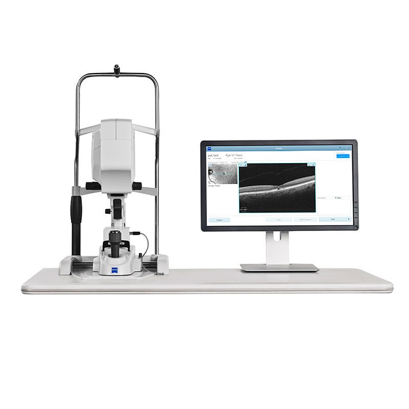
Reveal the hidden structures in the retina with the new PRIMUS 200 from ZEISS, the pioneer in ophthalmic OCT. Ideal for small practices, PRIMUS 200 delivers the essential clinical applications to help you manage your patients with certainty.
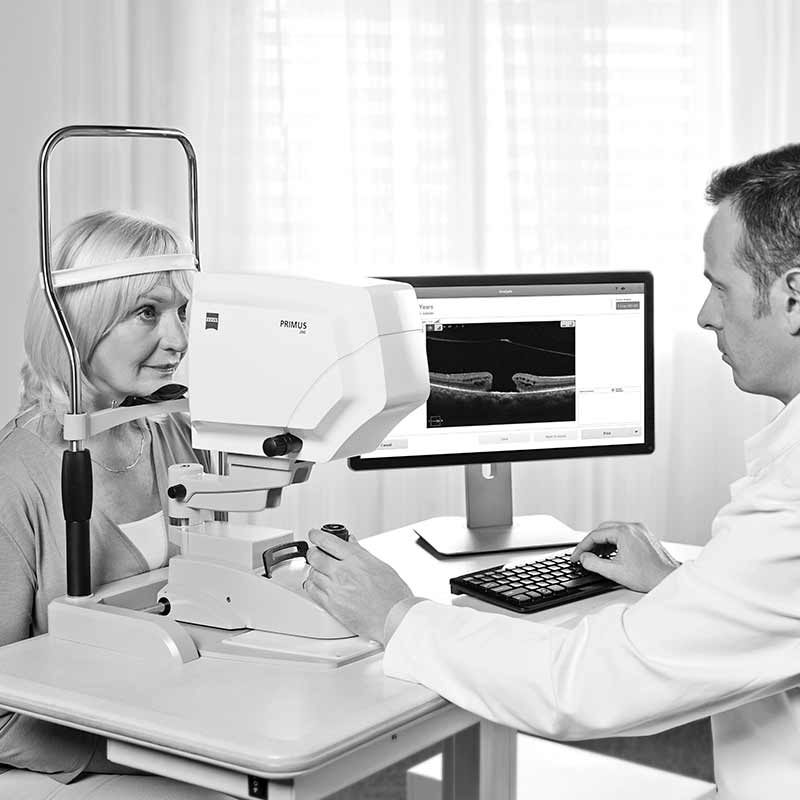
ZEISS PRIMUS 200 is the technology engine with the power and performance you need to deliver the optimal level of clinical care and to grow your practice.
Today, optical coherence tomography (OCT) has become as essential to clinical eye care as perimetry or fundus photography. OCT gives clinical insight to better understand your patient’s condition and the power to manage a broader range of pathologies within your practice.
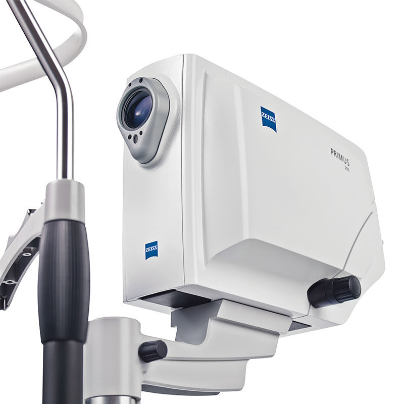
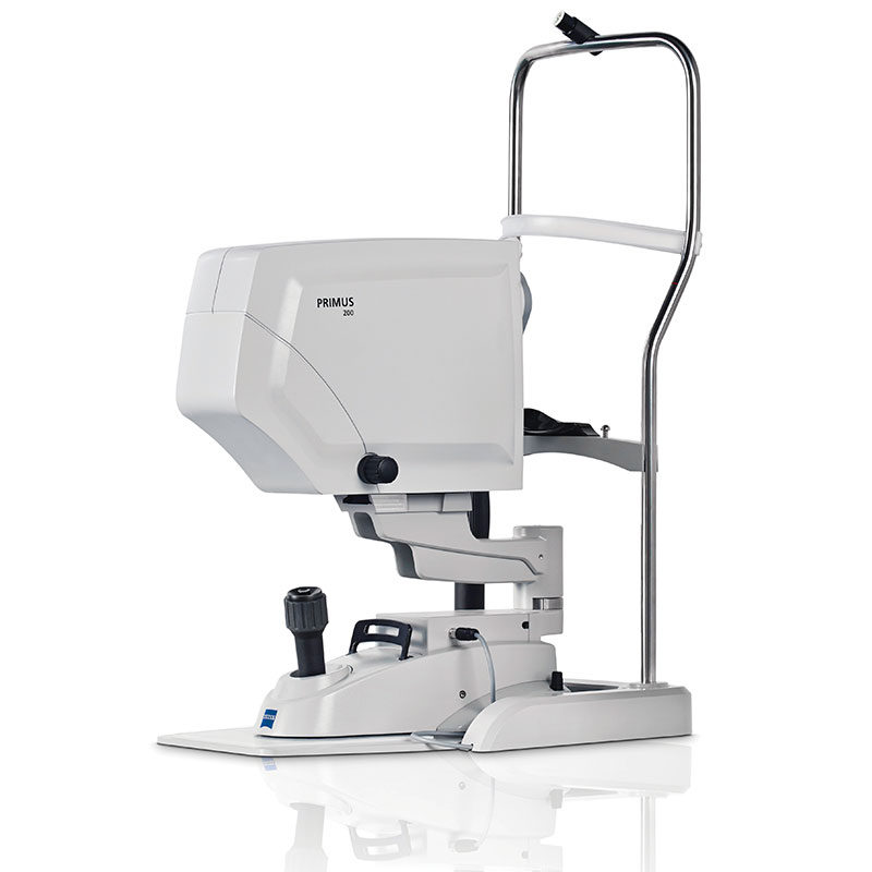
By incorporating legendary ZEISS optics and proprietary algorithms, in a compact and intuitive design, the ZEISS PRIMUS 200 is both a diagnostic and a patient education tool as it helps you to effectively communicate the essential aspects of a comprehensive treatment plan.
Primus 200 is the ideal OCT for small private practices.
Simple and intuitive in its design, Primus 200 helps you elevate your practice with:
Clearer images for deeper insight ZEISS PRIMUS 200 inherits the fundamental quality of world-class ZEISS optics, providing you with clear and compelling OCT and fundus images to help you detect pathologies at the earliest possible moment.
- A broader range of services
- An enhanced patient experience
- Optimized practice workflow for you and your staff
Manage more patients
- Manage a broader range of diseases
- Visualize retinal conditions with high-definition imaging
- Identity and monitor at-risk patients for glaucoma with comprehensive reports
Increase practice efficiency
- Simple 3-step smart workflow to minimize patient chair time
- Intuitive for operators at any level
- Advanced gold-standard OCT technology in an elegant, compact design
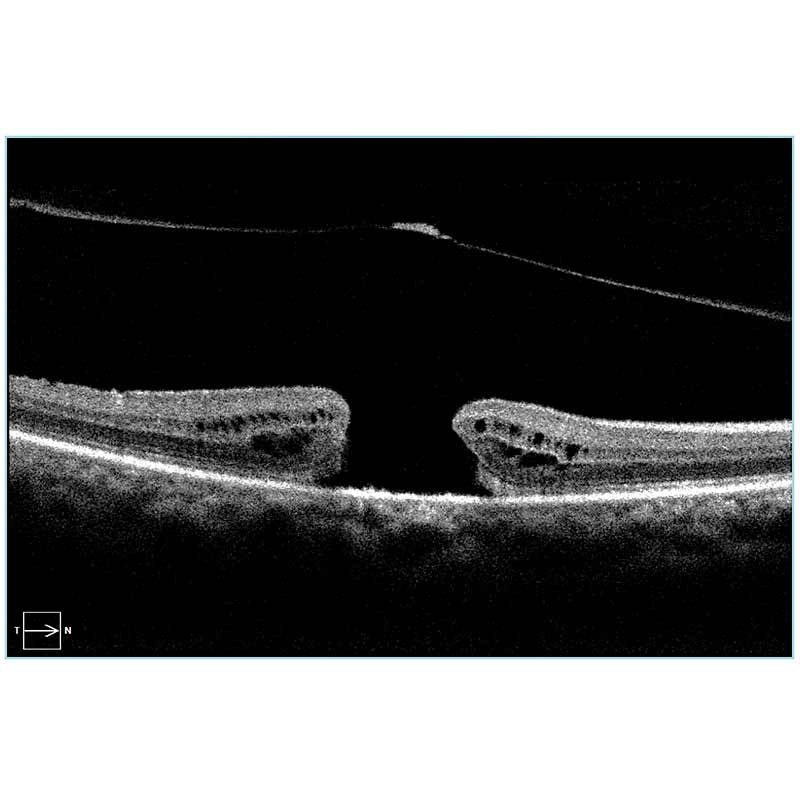
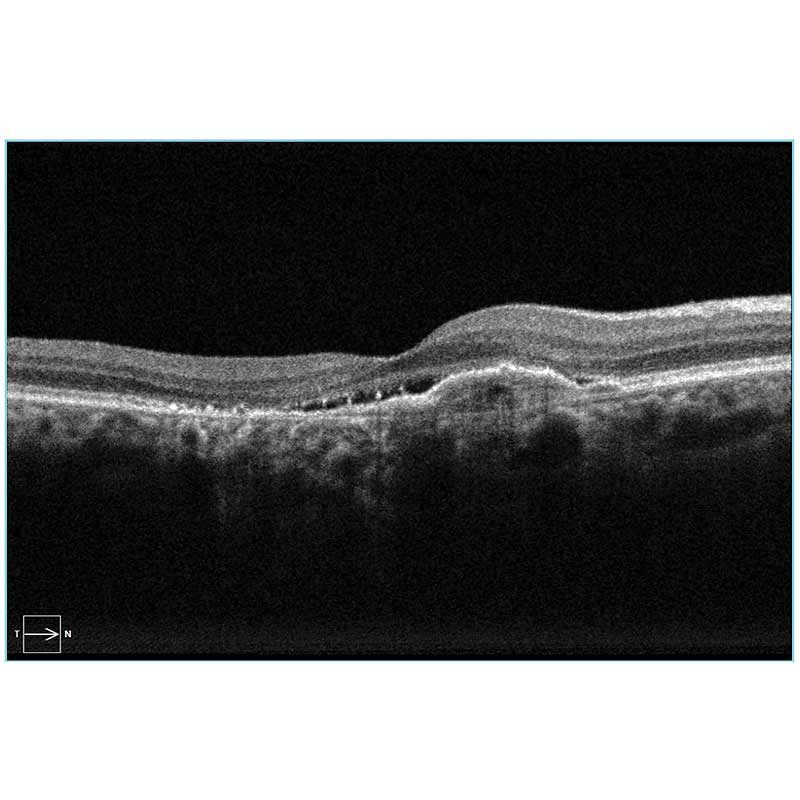
High-Definition B-scans and fundus imaging
PRIMUS 200 high resolutions B-scans reveal critical details to help you effectively manage a broad range of diseases.
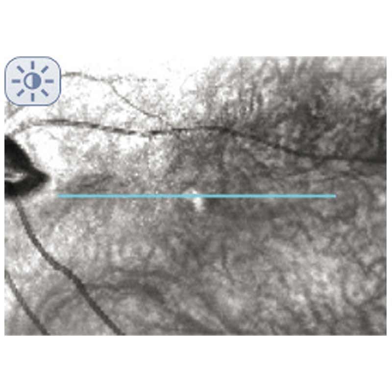
Clear sharp fundus images confirm the B-scan placement on the retina.
Minimal chin-time
Wide refraction range allows you to scan high myopia patients and increase patient throughput
ability to scan as small as 2mm pupil with no dilation, helps with patient comfort.
Operator-independent scan placement
Auto FoveaFinder™ and AutoCenter™ let operators of any skill level correctly position the scans on the fovea or optic nerve head. This ensured optimal repeatability on RNFL and ONH measures as well as allows visit-to-visit comparison of B-scans taken at the same.
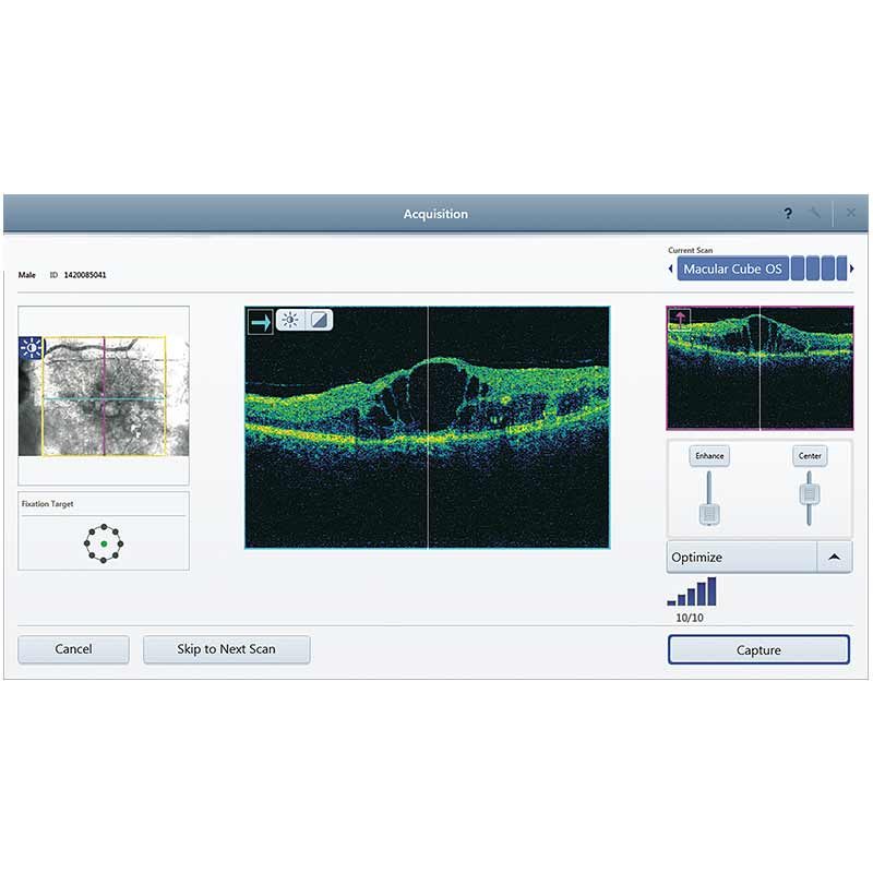
Report Driven Navigation
Simple 3-Step Smart Workflow
Select: Report Driven Workflow enables selection of desired reports to automatically generate the required scan-protocols for acquisition.
Capture: System-guided navigation leads you through easy acquisition protocols, and helps optimize scan quality with real time signal strength feedback.
Review: Pre-selected reports are automatically prepared for you to scan and select with the click of a button.
| Methodology | Spectral domain OCT |
| Optical source | Superluminescent diode (SLD), 840 nm |
| Scan speed | 100,000 scans per second |
| A-scan depth | 2.0 – 2.9mm (in tissue) |
| Axial resolution | 5 μm (in tissue) |
| Transverse resolution | 15 μm (in tissue) |
| Fixation | Internal, external |
| Internal Fixation (focus adjustment) | -20D to +20D (diopters) |
| Imaging Modes | Posterior segment, Anterior Segment, OCT Angiography, Fundus Imaging |
| B-scan | 12 mm Raster; 3-12 mm cube |
| OCTA | 3x3mm, 6x6mm, 8x8mm, 12x12mm (Retina); 4.5×4.5mm (Optic Nerve Head), 14×14 mm, 14×10 mm |
| Instrument Weight | 35 kg (77 lbs) (without monitor) |
| Instrument Dimensions (WxDxH) | 62.2L x 42.5W x 29.2H cm (24.4L x 16.7W x 11.4H in) |
| Monitor | 22” Widescreen HD |
| Input devices | wireless keyboard, wireless mouse |
| Operating system (Instrument) | Windows® 10 |
| Processor | i7 Intel® processor (7th gen) |