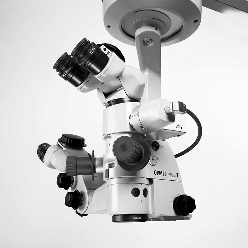
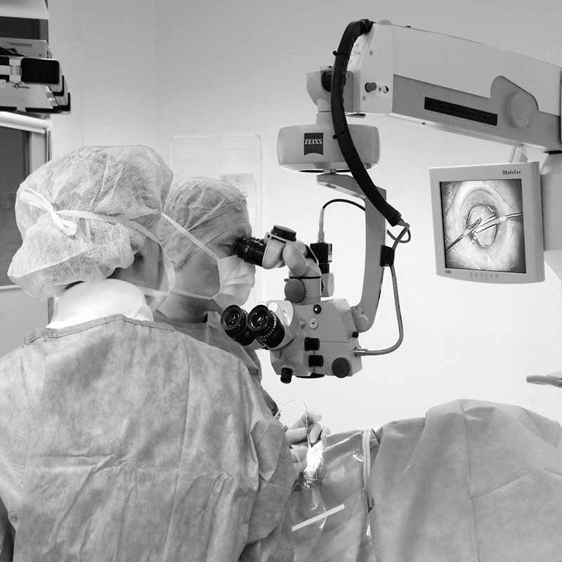
See the details of the eye with Zeiss Lumera T
Instant red reflex brightly illuminates the eye – due to Stereo Coaxial Illumination (SCI) –even with mature cataracts thanks to Stereo Coaxial Illumination (SCI)
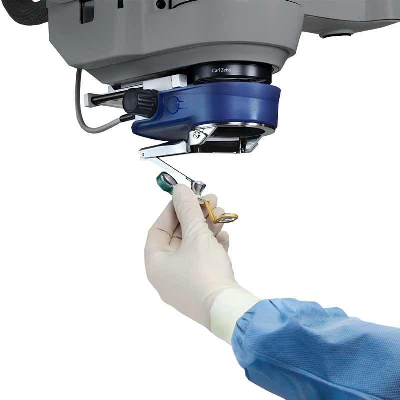
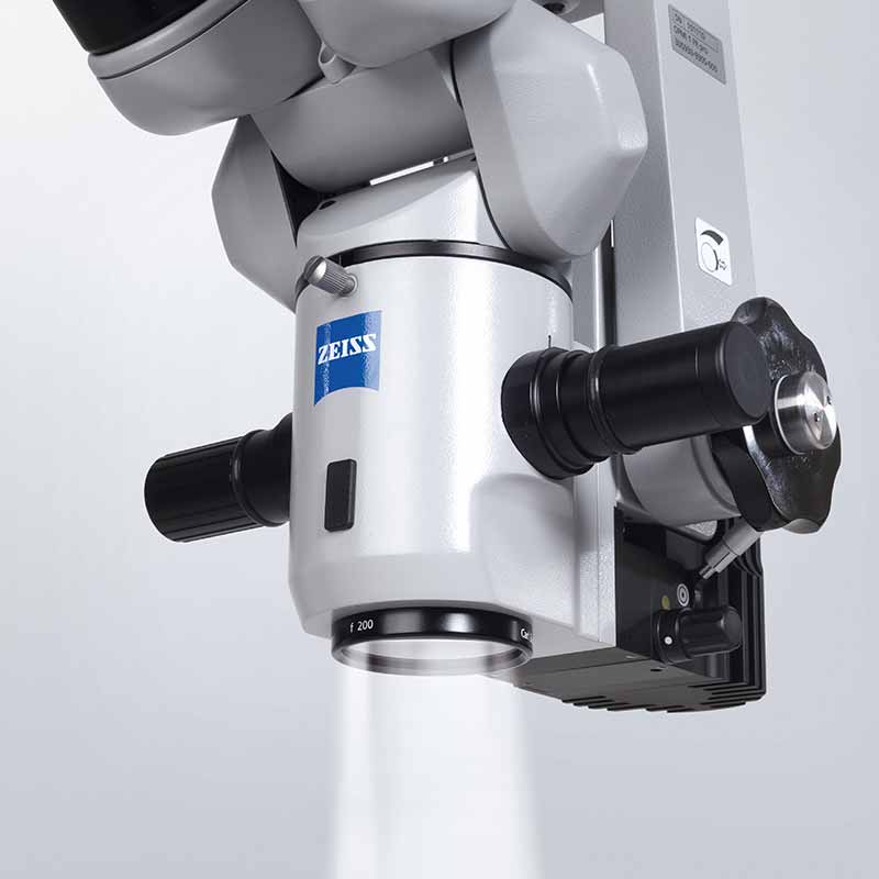
Integrated Superlux® Eye xenon illumination allows you to view the structure of the eye in great detail.
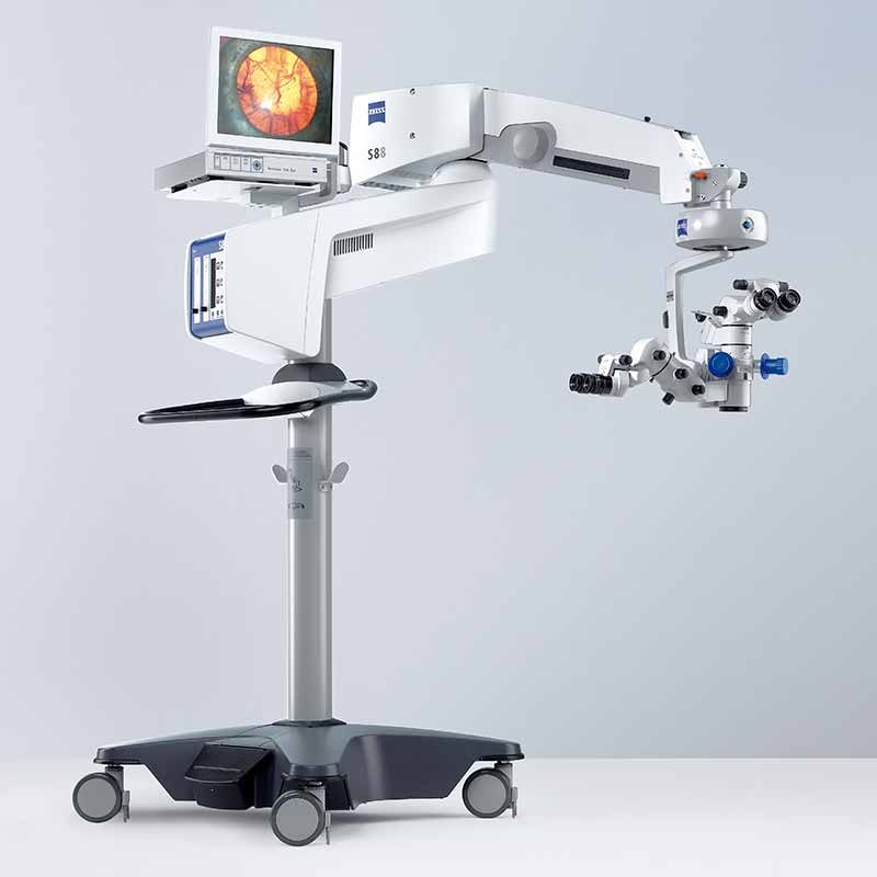
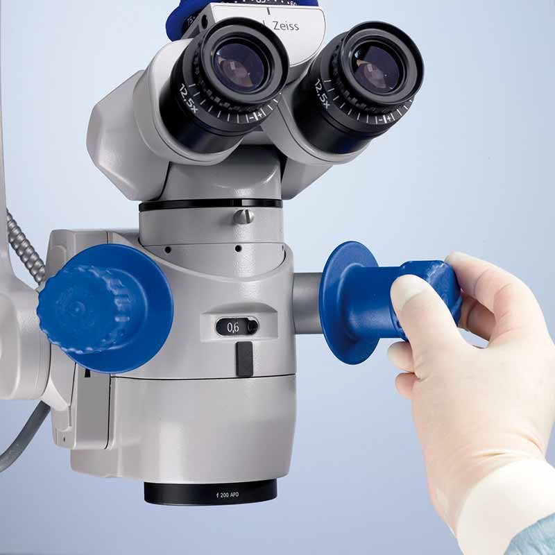
Effortless positioning with magnetic brakes
When the brakes are released the system smoothly glides into a new position. When locked, the surgical microscope remains firmly in place.
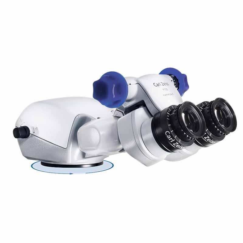
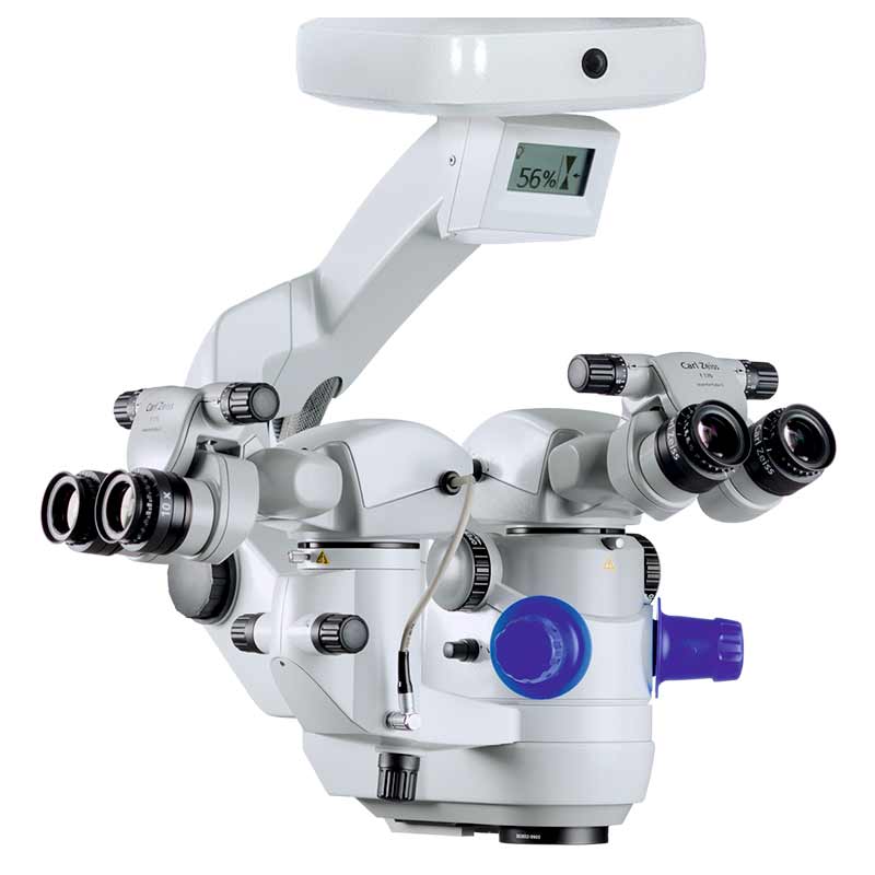
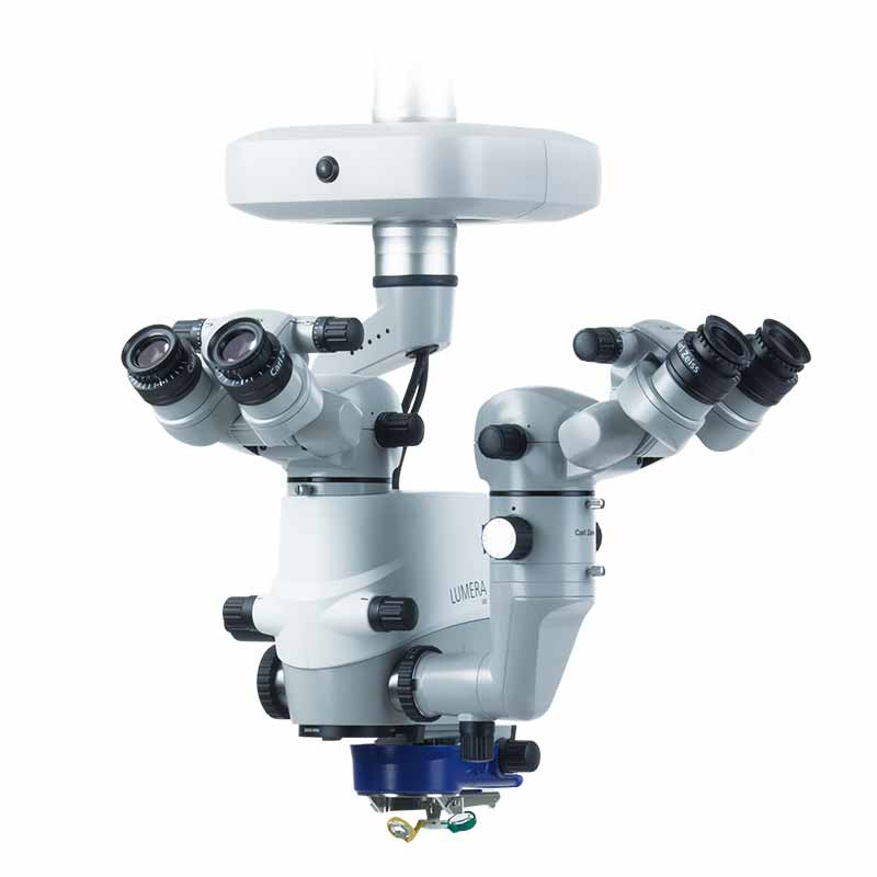
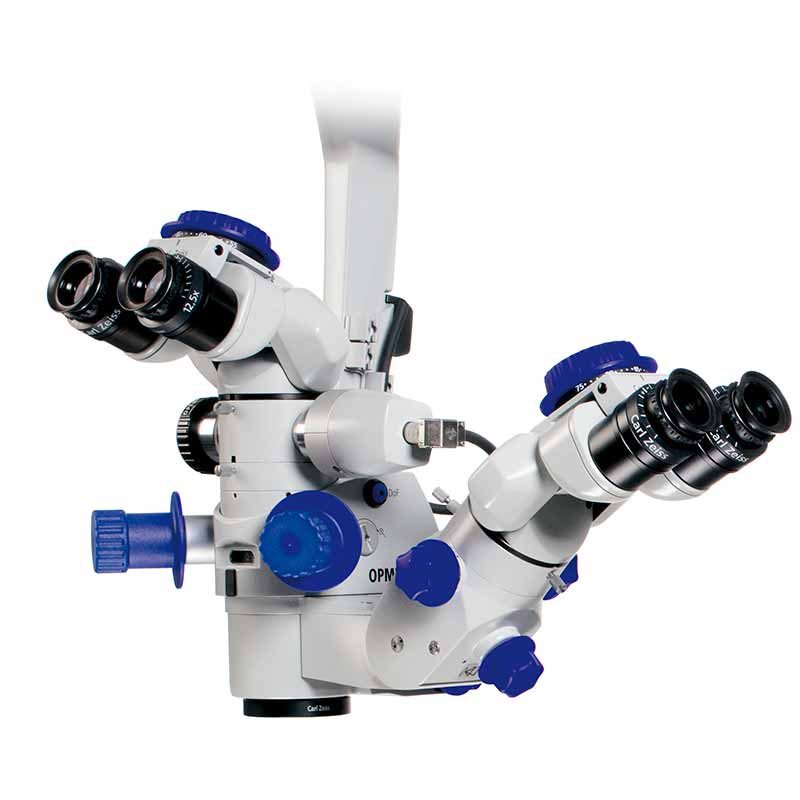
Independent second view
The ZEISS Lumera T surgical microscope can be equipped with a completely integrated assistant’s microscope. The second surgeon selects the focus and magnification independently of the main surgeon, thus enabling active assistance.
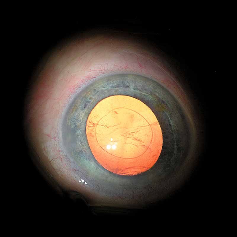
Natural Color Impression
The integrated Superlux® Eye xenon illumination allows you to see the anatomic structure of the eye in its natural colors and highly accurate detail. The use of the HaMode™ filter allows surgeons who prefer halogen to quickly switch to a light spectrum equivalent to halogen. This is particularly beneficial when several surgeons with different preferences regarding the light source use one system.
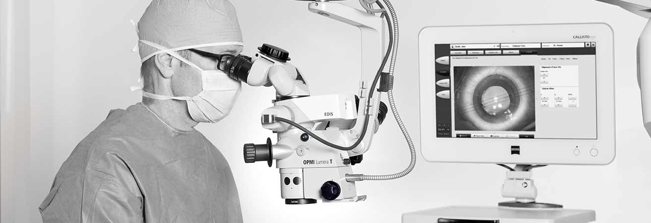
External Video Components
To support all requirements or wishes for customized video solutions, external components can be mounted to the surgical microscope system. The external attachment via standard optical and mechanical interfaces increases the flexibility and retrofitability of the surgical microscopes.
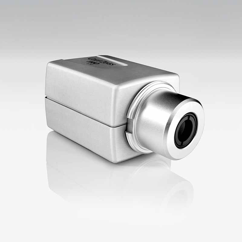
Video Cameras
TRIO 610 with CCU TRIO 600 – 3 Chip HD Camera System:
The high definition camera system with apochromatic video optics allows surgical microscope images to be generated with enhanced resolution and color rendition. The camera can be used for information, documentation, teaching and presentation of high quality images.
MediLive Trio Eye – 3 Chip SD Camera System:
Standard definition video camera with high sensitivity especially designed for different light conditions in anterior and posterior segment. The video images can be used for information, teaching, and documentation purposes. The MediLive® Trio Eye helps the surgeon to easily overcome the challenges of ophthalmic imaging.
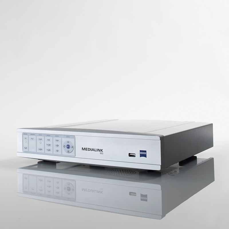
MEDIALINK 100:
Standard definition video recorder with image data management, that enables surgeons to record videos and capture still images. Using the MEDIALINK™ 100 standard definition videos and images can be automatically transferred to USB storage media or file servers.</p
HD Video Recorder:
A medical grade high definition video recorder is available.
MediLive Trio Eye – 3 Chip SD Camera System:
Standard definition video camera with high sensitivity especially designed for different light conditions in anterior and posterior segment. The video images can be used for information, teaching, and documentation purposes. The MediLive® Trio Eye helps the surgeon to easily overcome the challenges of ophthalmic imaging.
MEDIALINK 100:
Standard definition video recorder with image data management, that enables surgeons to record videos and capture still images. Using the MEDIALINK™ 100 standard definition videos and images can be automatically transferred to USB storage media or file servers.
HD Video Recorder:
A medical grade high definition video recorder is available.
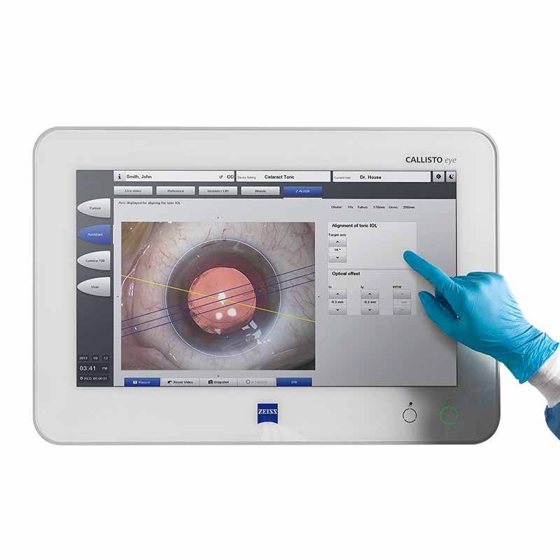
HD Video Monitors
Medical grade HD video monitors (1080p) are available in different sizes that match the surgical microscope video solutions.